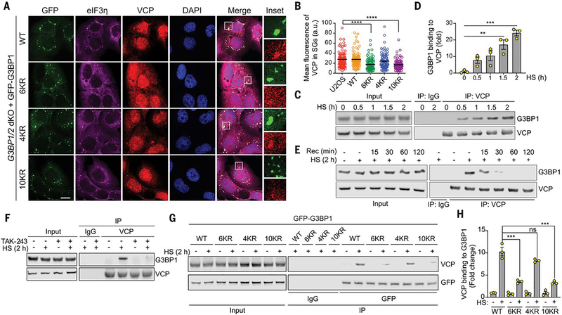
Fig. 3. VCP interacts with ubiquitinated G3BP1 in response to heat shock.
(A) Fluorescent imaging of U2OS G3BP1/2 dKO cells stably expressing GFP-G3BP WT, 6KR, 4KR or 10KR exposed to heat shock for 1 hour. Scale bar, 20 μm. (B) Fluorescence intensities of VCP in eIF3η-positive stress granules from three technical replicates (n = 90). Error bars indicate SEM. ****P < 0.0001 (ANOVA with Tukey’s test). (C) Immunoblot of U2OS cell extracts exposed to heat shock for the indicated times. Cell extracts were captured with magnetic beads conjugated with VCP antibody for IP, and resulting beads were analyzed by immunoblot. (D) Quantification of immunoblots from three biological replicates. Error bars indicate SEM. **P < 0.01, ***P < 0.001 (ANOVA with Tukey’s test). (E) Immunoblot showing G3BP1-VCP interaction in U2OS cells based on duration of recovery after heat shock for 2 hours. Cell extracts were captured with magnetic beads conjugated with antibody to VCP for IP, and the resulting beads were analyzed by immunoblot. (F) Immunoblot of U2OS cells treated with DMSO or TAK-243 (60 min) and exposed to heat shock for 2 hours. Cell extracts were captured with magnetic beads conjugated with antibody to VCP for IP. Blots show an attenuated G3BP1-VCP interaction with inhibition of ubiquitination. (G) Immunoblot of U2OS G3BP1/2 dKO cells stably expressing G3BP1 WT, 6KR, 4KR, or 10KR and exposed to heat shock for 2 hours. Cell extracts were captured with magnetic beads conjugated with antibody to GFP for IP. (H) Quantification of immunoblots from three biological replicates. Error bars indicate SEM. ***P < 0.001 (ANOVA with Tukey’s test).

