Figure 1.
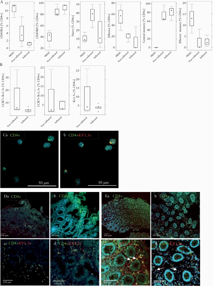
Kv1.3 expression on T cells in PBMCs and colonic biopsies from IBD patients. [A] Distribution of subtypes of antigen experienced and inexperienced CD4+ T cells in PBMCs and colonic biopsies from IBD patients [n = 3]. Isolated leukocytes were subjected to flow cytometric analysis and frequencies of subtypes of CD4+ cells in isolated PBMCs, and inflamed and non-inflamed areas of the colon are depicted as boxplots. [B] Frequencies of Kv1.3-expressing effector and naïve/central memory cells in biopsies of IBD patients [n = 6] in inflamed and non-inflamed [n = 5] areas depicted as boxplots. Boxes represent upper and lower quartiles, whiskers represent variability, and outliers are plotted as individual points. [C] Qualitative IHC of CD4+ T cells expressing Kv1.3 in PBMCs. [a] CD4; [b] CD4 and Kv1.3. Qualitative IHC of colonic biopsies from UC [D, n = 1] and CD [E, n = 1] patients. [a] CD4, non-inflammatory region. [b] CD4, inflammatory region, [c] CD4 and Kv1.3, non-inflammatory region, [d] CD4 and Kv1.3, inflammatory region. CD4 was stained with Alexa Fluor 433 [green] and Kv1.3 with Alexa Fluor 647 [red], cell nuclei with Dapi [blue]. PBMC, peripheral blood mononuclear cells; IBD, inflammatory bowel disease; UC,ulcerative colitis; CD, Crohn’s disease.

