
FIGURE 2.
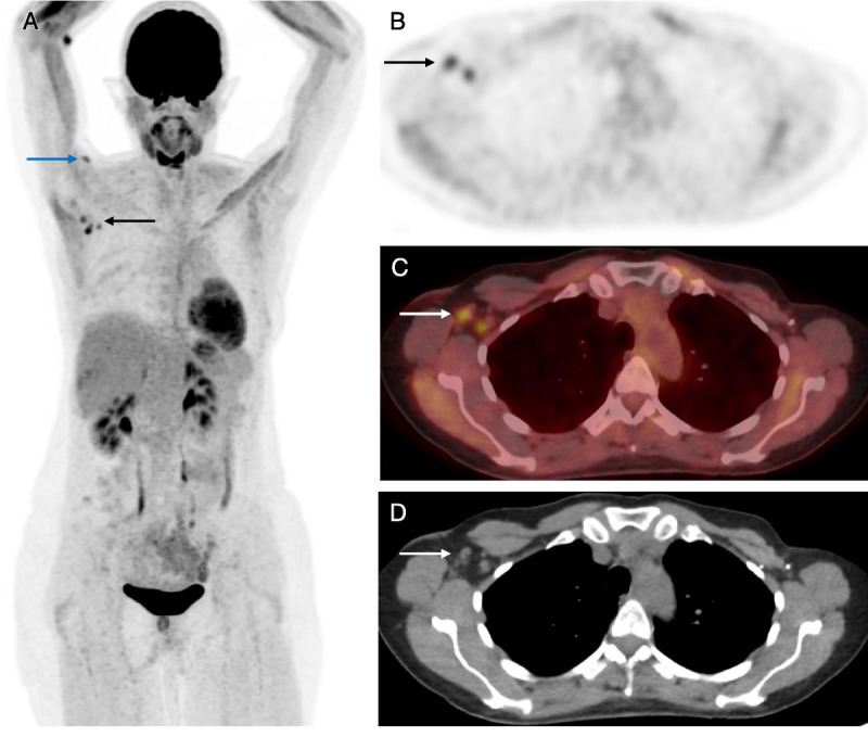
A 48-year-old woman with a history of primary left-sided breast cancer diagnosed 7 years previously and treated with left mastectomy and axillary node clearance plus risk-reducing right mastectomy and bilateral reconstruction presents for restaging scan. 18F-FDG PET/CT images (A, MIP; B, axial PET; C, axial fused PET/CT; D, axial CT) demonstrated intense uptake in nonenlarged right axillary lymph nodes (A–D, black and white arrows). The patient received the COVID-19 vaccine in the right arm 3 weeks earlier. There is a small focus of FDG avidity in the deltoid muscle in the right upper arm at the known injection site (blue arrow). This patient also underwent ultrasound-guided biopsy of the lymph node, which was shown to be reactive with no evidence of malignancy. At this time, the UK vaccination program was ahead of most other countries in the world, having offered the first dose of the vaccine to all over 70s by mid-February 2021. Being largely similar in age demographic, it became apparent that we would be seeing an increasing number of recently vaccinated patients presenting for surveillance cancer imaging. A new approach was needed to prevent unnecessary interventions, and our institution began to collect information about vaccination such as date, type, first/second dose, and arm vaccinated before PET/CT studies to avoid this pitfall in future patients.