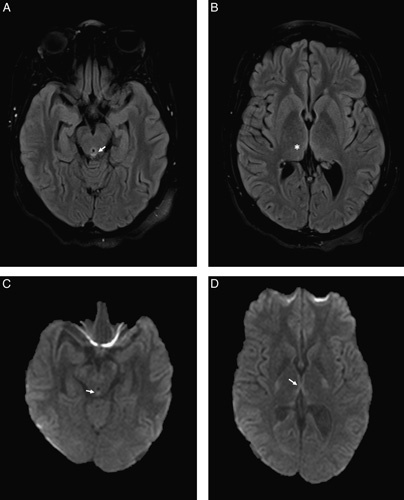
FIGURE 1.

MRI brain revealed findings suggestive of Wernicke encephalopathy with hyperintense lesions on FLAIR sequences in the periaqueductal gray and dorsomedial thalami (A, B) and restricted diffusion on DWI sequences in the periaqueductal gray and around the third ventricle (C, D). The white arrows illustrate the changes in the periaqueductal gray area and the star illustrates the changes in the dorsomedial thalami seen in Wernicke Encephalopathy on MRI. DWI indicates diffusion-weighted imaging; FLAIR, fluid-attenuated inversion recovery; MRI, magnetic resonance imaging.
