
Figure 3: Endothelial-mesenchymal transition signature in kidneys from HFpEF mice.
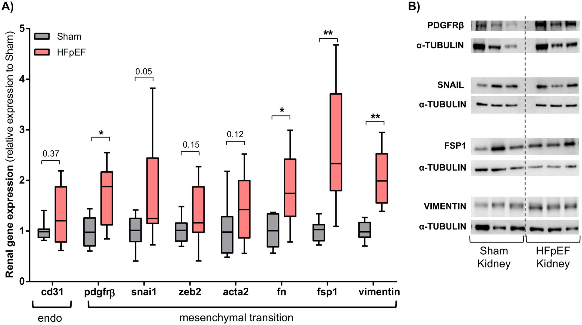
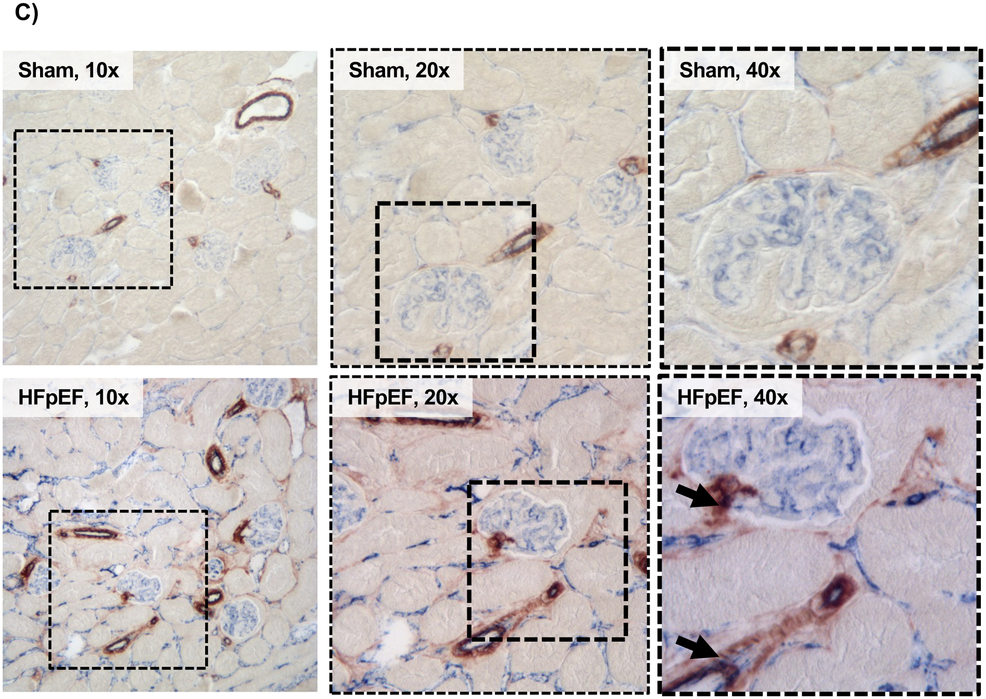
(A) Gene expression analysis of cluster of differentiation 31 (cd31), platelet-derived growth factor receptor beta (pdgfrβ), snail (snai1), zinc finger E-box-binding homeobox 2 (zeb2), smooth muscle actin alpha 2 (acta2), fibronectin (fn), fibroblast-specific protein 1 (fsp1) and vimentin in the kidneys of Sham and HFpEF mice. n = 8–9/group (B) Representative blots of PDGFRβ, snail, FSP1 and vimentin. (C) Renal immunostaining of CD31 and smooth muscle actin alpha 2 (ACTA2) in Sham and HFpEF mice, showing CD31 (blue) and ACTA2 (brown) staining but only in HFpEF were there double-stained cells (black arrows; bottom panel, 40X). Squared area shows magnification. Statistical analysis by 2-tailed Student t test for normally distributed data or Mann-Whitney U for those variables that were non-normally distributed. *P<0.01; **P<0.001.