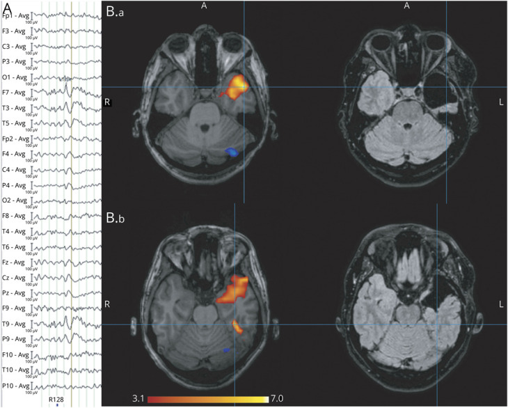
Figure 1. Example of a Concordant Study.

In this bilateral temporal case, left temporal interictal epileptiform discharges (n = 32) consisting of spikes with maximum amplitude at F7, T3,T9, and P9 followed by a slow wave (A) resulted in an activation cluster in the left anterior temporal lobe. Preoperative and postoperative MRIs. (B.a) The primary cluster with a peak value of t = 7.39 was included in the resection. (B.b) The next most significant cluster with a peak value of t = 6.38 was an activation cluster in the left fusiform gyrus, which was not resected. These t values are above false discovery rate level (4.72). This case fulfilled our first 2 levels of confidence but did not fulfill the criterion for high confidence results because the t value of the second most significant cluster was close to the first. This example would be predicted as a good outcome case with medium confidence, but this patient belongs to the poor outcome group (Engel 3).
