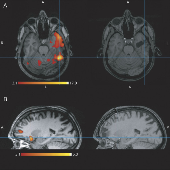
Figure 3. Two Examples of Partially Discordant Studies.

(A) The peak (t = 17.41) of the primary cluster in the left fusiform gyrus was outside the resection. The resection includes part of the same cluster in the anterior temporal lobe, but because the peak is >2 cm from the resection borders, it is not considered partially concordant. In this case, the second most significant cluster had peak t = 6.20 (not shown), so it fulfills the criterion for high confidence results and would be predicted as a poor outcome case with high confidence. The actual outcome was indeed Engel class 4. (B) The primary cluster with peak t = 4.99 in the right orbitofrontal area is not included in the resection. There is another activation cluster (peak t = 3.88) in the resection cavity at the frontal pole; therefore, the results are partially discordant. These clusters are all below the false discovery rate (5.00) level, resulting in a low confidence study. In this case, the prediction would be poor outcome with low confidence. The patient improved, having only rare nocturnal events and no diurnal ones (Engel class 2).
