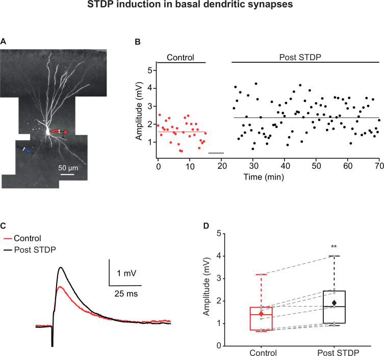
Figure 2. Long-term potentiation (LTP) induction via spike timing-dependent plasticity (STDP) protocol in basal dendrites of piriform cortex (PCx) pyramidal neurons.
(A) A layer IIa pyramidal neuron filled with CF633 (200 µM) and OGB-1 (200 µM) via the somatic recording electrode (red electrode). Focal stimulation was performed using a double-barrel theta electrode located nearby a distal basal dendritic site (blue electrode; 129.25 ± 8.83 µm from soma). (B) Amplitude of single EPSPs is represented over time for control stimulation and after STDP induction protocol (gray bar represents the time of induction stimulus). Induction protocol, as described in Figure 1B. Bottom: traces of average EPSPs from the cell shown in (A) for control (red) and post induction (black). (C) Traces of average EPSPs evoked during control (red) and after STDP induction (black). (D) Box plot showing EPSP amplitudes pre- and post-STDP induction during control (red), and post-STDP induction (black) for distal basal input stimulation. EPSPs were significantly enhanced post-induction protocol (137.12% ± 6.11% of the control; p=0.0031, n = 8). The gray dots represent the average EPSP for each cell, and the diamond represents the mean EPSP of the entire set. Dotted gray lines connect between pairs of control and post-induction values.

