. 2021 Oct 26;10:e70383. doi: 10.7554/eLife.70383

Figure 3. Long-term potentiation (LTP) of lateral olfactory tract (LOT) inputs by NMDA-spikes in piriform cortex (PCx) pyramidal neurons.

(A) Fluorescence image of a layer IIa pyramidal neuron filled with CF633 (200 µM) and OGB-1 (200 µM) via the patch recording electrode (red electrode). A focal synaptic stimulating theta electrode was placed at the distal apical dendrite at layer Ia (blue electrode; 272.75 ± 6.6 µm from the soma). (B) Voltage responses evoked by gradually increasing synaptic stimulation intensity (burst of three pulses at 50 Hz). Peak voltage response as a function of stimulus intensity showing an all-or-none response (left). (C) Schematic of NMDA-spike induction protocol (bottom). NMDA-spikes evoked by three pulses at 50 Hz, repeated at 4 Hz for 2–7 times. Upper panel: example voltage response to NMDA-spikes induction protocol. (D) Amplitude of single EPSPs is represented over time for control stimulation (red), after STDP induction protocol (black) and after NMDA-spike induction protocol (blue). Gray bars represent the time of induction stimulus. Control EPSPs were recorded at 0.033 Hz for 10–15 min. Potentiation was observed only after the NMDA-spike induction protocol. Bottom: traces of average EPSPs in control (red), post-STDP (black), and post-NMDA-spike induction (blue) for the cell shown in (A). (E) Plot of % LTP (relative to control EPSPs) vs. number of NMDA-spikes evoked during the induction protocol. All values are insignificant. (F) Box plot showing the EPSP amplitude during control (red) and post-NMDA-spike induction protocol (blue). NMDA-spike induction protocol induced large potentiation of the control EPSP (213.98% ± 10.81%; p=0.0000687; n = 26). (G) Box plot showing the EPSP amplitude during control NMDA-spike induction protocol (red) and after induction in the presence of APV (50 µM; green). No significant change in EPSP was observed when NMDA-spikes were blocked with APV (95.06% ± 4.69% of control; p=0.817, n = 5). (H) Box plot showing the EPSP amplitude for control NMDA-spike induction protocol (red) and after induction with sub-NMDA EPSPs (teal). No significant change in EPSP amplitudes was observed (106.79% ± 5.69%, p=0.8373, n = 5). In box plots, the gray dots represent the average EPSP for each cell, and the diamond represents the mean EPSP of the entire set. Dotted gray lines connect between pairs of control and post-induction values. See also Figure 3—figure supplements 1 and 2.

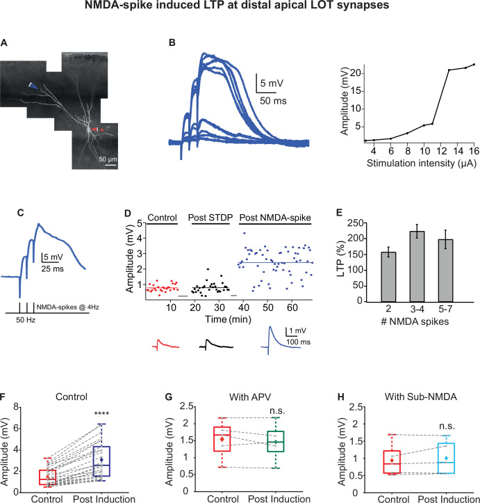
Figure 3—source data 1. Data for Figure 3B, D, F-H.

Figure 3.

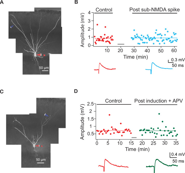
Figure 3—figure supplement 1. Example experiments showing NMDA-spikes are necessary for long-term potentiation (LTP) of lateral olfactory tract (LOT) synapses.

Figure 3—figure supplement 1.

(A) Reconstruction of a layer IIa pyramidal neuron filled with CF633 (200 µM) and OGB-1 (200 µM) via the somatic recording electrode (red) and focally stimulated using a theta electrode in distal apical dendrite at layer Ia (blue; 298 ± 14.38 µm from soma). (B) Amplitude of single EPSPs is represented over time for control stimulation (red) and after EPSPs subthreshold to NMDA-spike induction protocol (teal). Same induction protocol as in Figure 3, except that stimulation was just subthreshold to NMDA-spikes initiation. Bottom panel: averaged EPSPs evoked during control (red) and post induction with sub-NMDA EPSPs (teal). (C) Reconstruction of a layer IIa pyramidal neuron filled with CF633 (200 µM) and OGB-1 (200 µM) via the somatic recording electrode (red) and focally stimulated using a theta electrode in distal apical dendrite at layer Ia (blue; 275.6 ± 11.55 µm from soma). (D) Amplitude of single EPSPs is represented over time for control stimulation (red) after NMDA-spike induction protocol in the presence of APV (50 µM; green). Same induction protocol as in Figure 3. Bottom panel: averaged EPSPs evoked during control (red) and post induction in the presence of APV (green).
Figure 3—figure supplement 1—source data 1. Data for Figure 3—figure supplement 1B and D.
Figure 3—figure supplement 2. Estimation of the number of synapses needed to initiate NMDA-spikes with in vivo-like odor stimulation.

Figure 3—figure supplement 2.

(A) The probability that at least a single branch will be simultaneously activated by 10 synapses or more during a typical odor stimulation. In this calculation, each pyramidal neuron (PN) contained 10 terminal apical branches, with a lateral olfactory tract (LOT) band of 100 µm and density of one synapse/1 µm yielding total 1000 LOT synapses/per neuron. For this calculation, a range of glomeruli inputs is explored, with each neuron randomly receiving 0.64 synapses from each glomerulus. (B) An example of the probability to randomly cluster inputs on at least a single branch given 110 activated glomeruli yielding a total of 70 synapses/neuron/odor (left) or when 200 activated glomeruli yielding 128 total active synapses/neuron/odor (right). For example, the probability to cluster 10 or more inputs (denoted by dashed line) on a single branch when a total of 70 or 128 inputs are activated was 15% or 80%, respectively.
Figure 3—figure supplement 2—source code 1. Source code for Figure 3—figure supplement 2 is provided.