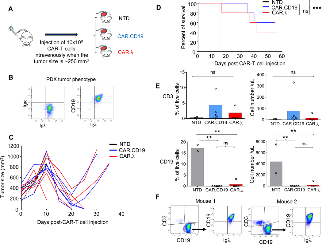
Figure 4. CAR.λ and CAR.CD19-T cells demonstrate equivalent antitumor effects in a patient derived xenograft (PDX) MCL model.
(A) Schema of PDX model in which MCL cells derived from a patient were injected in the flank of NSG mice. After tumor engraftment (tumor size of 250 mm3), mice were injected intravenously with either NTD, CAR.CD19, or CAR.λ-T cells. (B) Flow cytometry plots showing CD19 and Igλ expression in PDX cells. (C) Kinetics of tumor growth in all treated mice described in (A). (D) Kaplan-Meier survival curves of mice treated as described in (A). *** p-value < 0.001, log-rank Mantel-Cox test (n = 5). (E) CD3+ (upper panels) and CD19+ (lower panels) cells in the peripheral blood of treated mice described in (A) (n = 5). ** p-value <0.01, one way ANOVA. (F) Representative flow cytometry plots showing the expression of CD19 and Igλ in PDX cells that recurred in two mice treated with CAR.λ-T cells showing no loss of the Igλ.

