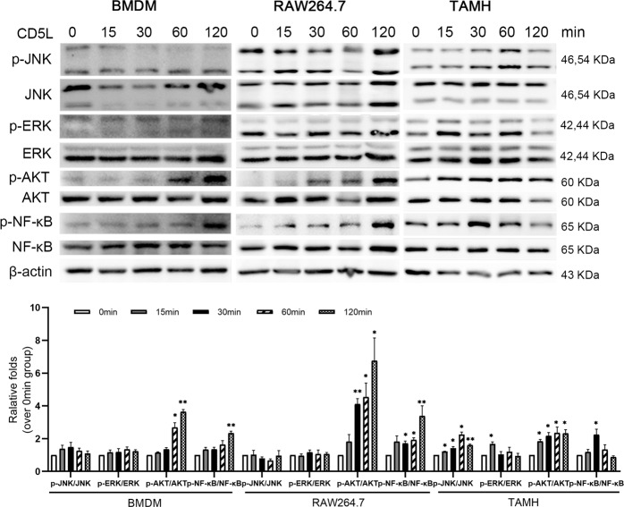
Fig. 3. CD5L protein activates mouse macrophages and hepatocytes in vitro.
CD5L protein (1 μg/ml) was co-incubate with BMDMs, RAW264.7 cells and TAMH cells, respectively. Cell proteins were collected at 0, 15, 30, 60, and 120 min, and the protein levels were detected by western blotting. The AKT, ERK, JNK, and NF-κB phosphorylation were evaluated using total ERK, NF-κB, AKT, and JNK as controls. The representative results from three independent experiments are shown and the data are presented as means ± SD. *P < 0.05; **P < 0.01.

