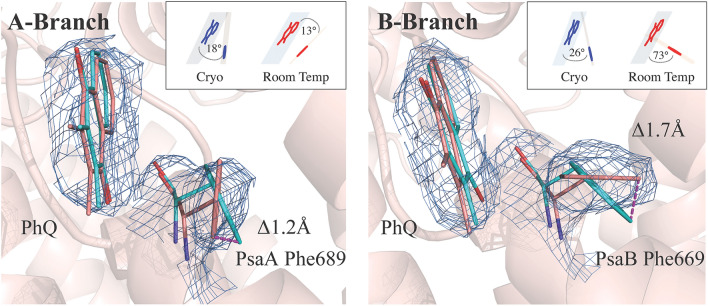
Figure 4.
2Fobs − Fcalc electron density contoured to 1.5 with the modeled phylloquinone in the PsaA and PsaB electron transport pathways. For aromatic residues PsaA-Trp697 and PsaA-Phe689 of PsaA and PsaB-Trp677 and PsaB-Phe669 of PsaB (salmon) the orientation obtained from the cryogenic structure (1JB0) is overlaid in cyan. PsaA-Phe689 is twisted relative to the cryogenic structure to a more parallel orientation with the phylloquinone. The B site reveals PsaB-Phe669 in an orientation more perpendicular to the plane of the phylloquinone. The stick representation of the PsaB-Phe669 and PsaA-Phe689 aromatic ring plane angles to phylloquinone in room temperature structure versus 1JB0 is shown in the inset. Both sites reveal significant shifts of atomic coordinates. Figure generated with Pymol v. 2.4.0a0. https://pymol.org.

