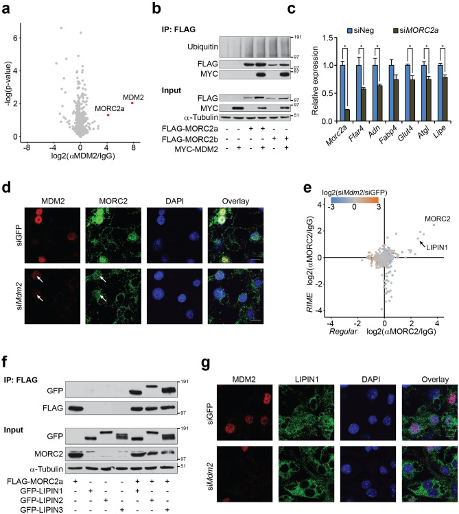
Figure 5.
MDM2 is necessary for nuclear localization of MORC2 and LIPIN1. (a) Volcano plot of proteins enriched by all three monoclonal antibodies directed against MDM2 in lysates from 3T3-L1 adipocytes. Plot generated in Perseus software70. (b) Coimmunoprecipitation against FLAG in lysates from cells ectopically expressing FLAG-MORC2s and/or MYC-MDM2. Western blot analysis against Ubiquitin, FLAG and MYC. α-Tubulin was included as loading control. (c) Real-time qPCR-based quantification of Morc2a, Ffar4, Adn, Fabp4, Glut4, Atgl, and Lipe in 3T3-L1 adipocytes with knockdown of MORC2a. (d) Localization of MDM2 and MORC2 in 3T3-L1 adipocytes with knockdown of Mdm2 as assessed by immunofluorescence. Nuclei were stained with DAPI. Grey scale bar corresponds to 10 µm. Images processed using ImageJ. (e) Scatterplot showing proteins enriched by MORC2 antibody with regular IP on the horizontal axis and RIME on the vertical axis. Colouring of dots refers to impact of Mdm2 knockdown with blue indicated decreased binding and orange augmented. Plots generated using Perseus software70. (f) Coimmunoprecipitation against FLAG in lysates from cells ectopically expressing FLAG-MORC2a and/or GFP-LIPIN1, − 2, or − 3. Western blot analysis against FLAG, GFP, and MORC2. α-Tubulin was included as loading control. (g) Localization of MDM2 and LIPIN1 in 3T3-L1 adipocytes with knockdown of MDM2 as assessed by immunofluorescence. Nuclei were stained with DAPI. Grey scale bar corresponds to 10 µm. Images processed using ImageJ. For a, p-value was calculated using Student’s t-test. For c, significance was tested using Student’s t-test, * = p-value < 0.05.

