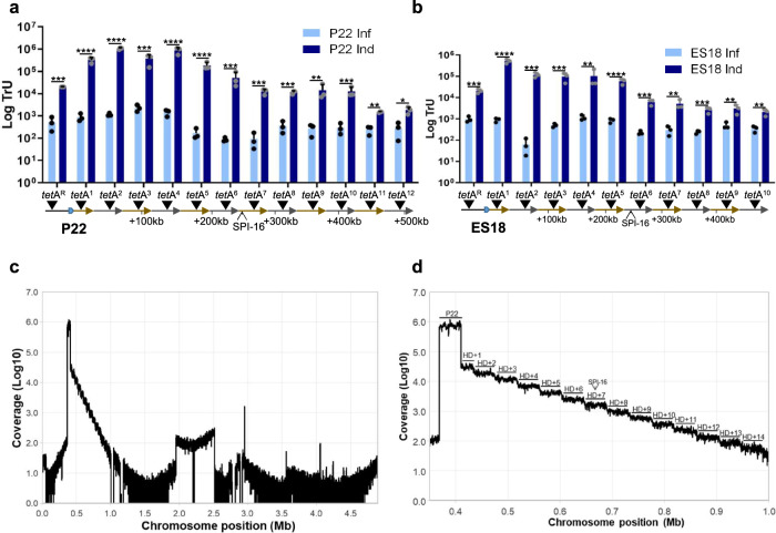
Fig. 4. P22 and ES18 engage in lateral transduction, transferring large metameric spans of the bacterial chromosome at high frequencies.
a The transfer of tetracycline (tetA) markers located upstream (tetAR) or downstream of the P22 attB site, in twelve successive capsid headfuls (tetA”n”), was tested following P22 prophage induction (Ind) or P22 infection (Inf). b The transfer of tetA markers located upstream (tetAR) or downstream of the P22 attB site, in ten successive capsid headfuls (tetA”n”), was tested after ES18 induction or ES18 infection. c A P22 lysogen was mitomycin C-induced and the resulting phage particles purified. The DNA from the phage particles was extracted and sequenced. The coverage of chromosomal DNA is represented. d Zoom of the region encompassing lateral transduction is visualized, displaying a successive ‘stepdown’ pattern in DNA packaging efficiency for each consecutive headful. Transduction units (TrU) per milliliter were normalized by PFU per milliliter and represented as the log TrU of an average phage titer (1 × 10⁹ PFU). Error bars indicate standard deviation from the mean of three independent experiments. For all panels, values are means (n = 3 independent samples). An unpaired two-sided t test was performed to compare mean differences between infection and induction in each marker. Adjusted p values were as follows: ns > 0.05; *p ≤ 0.05; **p ≤ 0.01; ***p ≤ 0.001; ****p ≤ 0.0001. The exact statistical values for each of the conditions tested are listed on Table S1.

