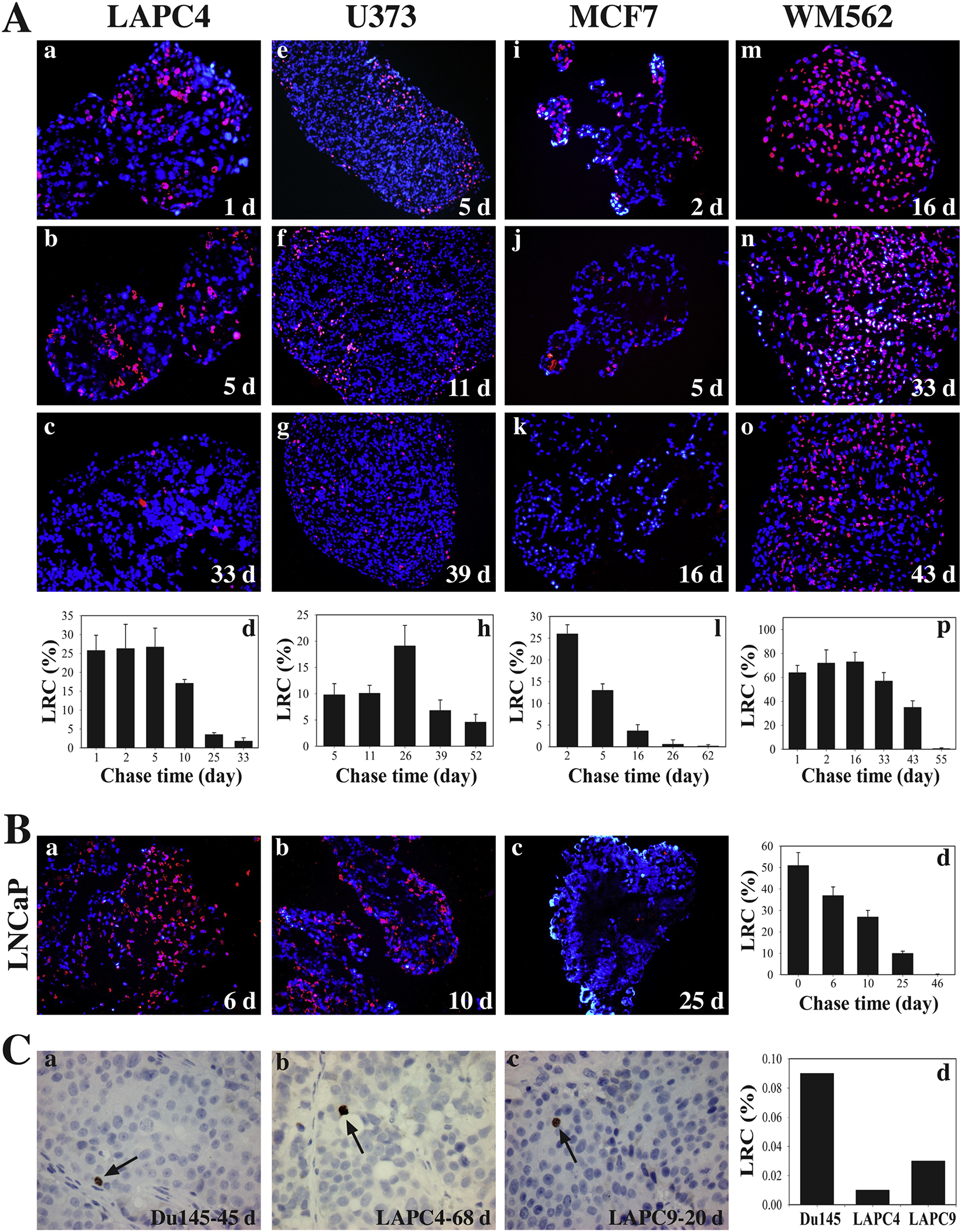
Fig. 4.

Human cancer cell spheres and prostate cancer xenografts harbor LT-LRCs (SCCs). (A-B) Human cancer cell spheres harbor LT-LRCs (i.e., SCCs) with distinct levels of quiescence. Human PCa (LAPC4 and LNCaP), glioblastoma (U373), breast cancer (MCF7) and melanoma (YVM562) cell-derived spheres were pulsed with 10 μM BrdU for 43 h, after which BrdU was washing off and sphere cultures chased for the days (d) indicated. The endpoint spheres were harvested and embedded in paraffin, and sections used in BrdU staining. At least 6 sections were analyzed and a total of 1,500 – 3,000 cells counted under an epifluorescence microscope for each cell type. The BrdU+ (i.e., label-retaining) cells were presented as bar graphs (mean ± S.D). (C) Human PCa xenografts have raie SCCs (LT-LRCs) in vivo. To identify LRCs in xenograft prostate tumors, 100 pi of BrdU solution (20 mM stock) was i.p injected (4x over 43 h) into the NOD/SCID mice bearing Du145, LAPC4, or LAPC9 xenograft tumors. Tumors were then “chased” for 45, 68 or 20 days (d), respectively, until harvest. Tumors were embedded in paraffin and serial sections of 5 pm were cut and used in immunohistochemical staining of BrdU. At least 6 sections were analyzed and counted for each tumor type and the BrdU ‘ cells scored under a microscope.
