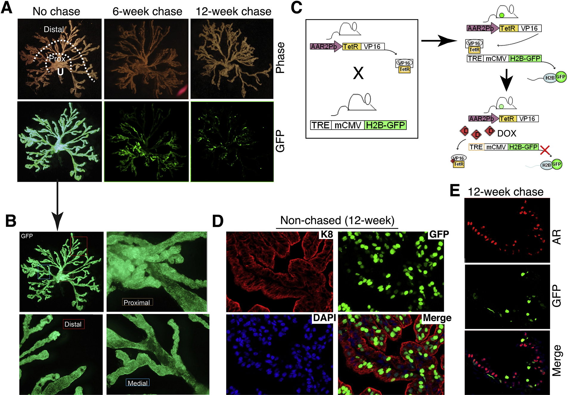
Fig. 6.

Adapting the inducible H2B-GFP labeling/chasing system to identify and study slow-cycling luminal progenitor cells in the mouse prostate. (A) Chase time-dependent decrease in GFP fluorescence intensity and in GFP+ cells. Shown on top are micro-dissected mouse prostate alveolar-ductal tree structure in relation to the urethra (U). Distal (secretory) alveoli and proximal (Prox) ducts close to the urethra are indicated. Shown below are the corresponding GFP images in non-chased (left; 20-week), 6-week chased (middle) and 12-week chased prostates. Note that the no-chase and 6-week chase images were adapted from [54] with permission. (B) The non-chased prostate in (A; the arrow) is shown in higher magnifications, to illustrate the homogeneous GFP labeling in the proximal, distal and medial regions of the mouse prostate. (C) Schematic of the model (see Text). (D) Analysis of a 12-week non-chased anterior prostate shows that the GFP+ cells are largely K8+ luminal epithelial cells. (E) Analysis of a 12-week chased ventral prostate reveals significantly reduced numbers of GFP+ cells, which are mostly AR−.
