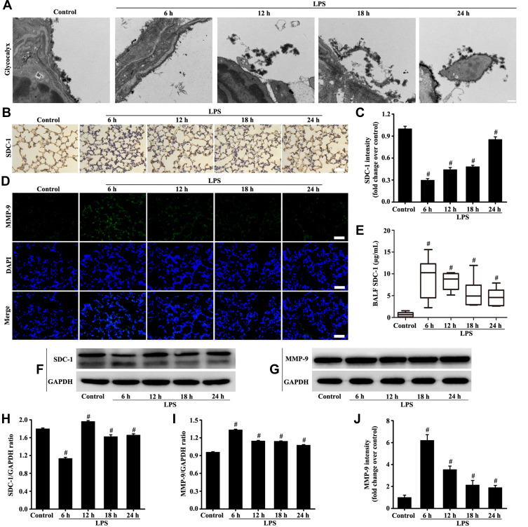
Figure 2.
Mice were intratracheally atomized with 5 mg/kg LPS (stimulation for 6, 12, 18, and 24 h) to observe the changes of glycocalyx and MMP-9. Glycocalyx was analyzed by an electron microscope (A, scale bar = 0.2 µm). SDC-1 was detected by immunohistochemistry (B, magnification, × 200; scale bar, 50 μm) and Western blot (F). The MMP-9 was detected by immunofluorescence (D, magnification, × 200; scale bar, 50 μm) and Western blot (G). The SDC-1 in the BALF was measured by SDC-1 ELISA (E). (C, H, and I) protein intensity analysis of (B, F, and G) respectively. (J) Fluorescence intensity analysis of (D). All data are presented as the mean ± SD of three independent experiments. #p < 0.05 vs the control group.

