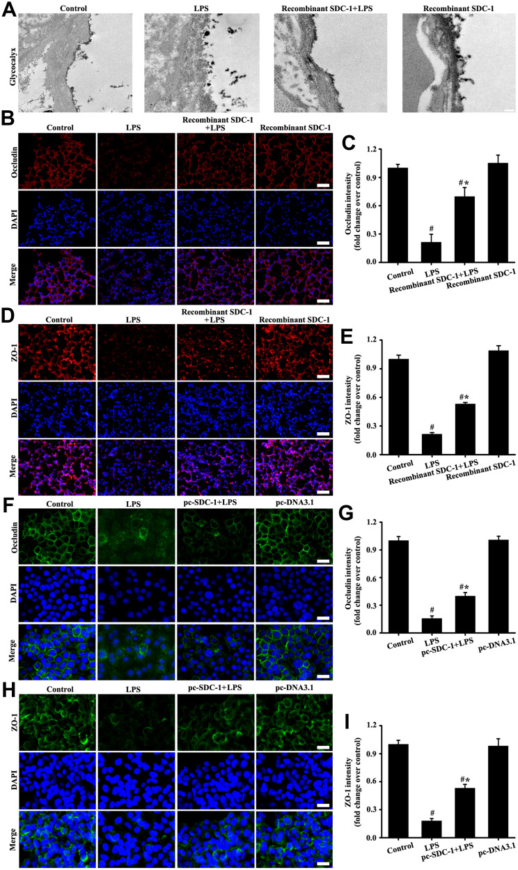
Figure 8.
Recombinant mouse SDC-1 protein alleviated glycocalyx shedding and tight junction damage in mice, and overexpression SDC-1 alleviated tight junction damage in A549 cells. The mice were intratracheally atomized with 500 ng/day recombinant SDC-1 protein for 7 days and then exposed to LPS to stimulate for six hours. The lipofectamine 3000 reagent and pc-SDC-1 (1 µg/mL) were employed for A549 cells transfection. Forty-eight hours after transfection, the A549 cells were cultured with LPS (100 ng/mL) stimulation for six hours. Glycocalyx was conducted by an electron microscope (A: scale bar = 0.2 µm). Occludin in mice (B, magnification, × 200; scale bar, 50 μm) and A549 cells (F, magnification, × 200; scale bar, 50 μm) were detected by immunofluorescence. The ZO-1 in mice (D, magnification, × 200; scale bar, 50 μm) and A549 cells (H, magnification, × 200; scale bar, 50 μm) were detected by immunofluorescence. (C, E, G, and I) are the fluorescence intensity analyses of (B, D, F and H) respectively. All data are presented as the mean ± SD of three independent experiments. #p < 0.05 vs the control group, #*p < 0.05 vs the LPS group.

