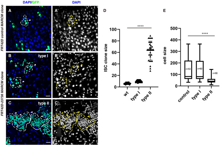
Fig. 1.
Two type of ISC clones are observed in FRT42D-23736 mutant. (A) FRT42D control ISC MARCM clones (green) (white arrowhead). One ISC MARCM clone is labeled with dotted lines. DAPI staining for the nucleus is showed separately. (B) Type I ISC MARCM clones from FRT42D-23736 mutant (green) (white arrowheads). Please note that the size of type I clone is larger than that of control clone. DAPI staining for the nucleus is showed separately. (C) Type II ISC MARCM clones from FRT42D-23736 mutant (green) (white arrowheads). Please note that the size of type II clone is much larger than that of control and type I clones and the cells in these clones are small and quite uniform in size. DAPI staining for the nucleus is showed separately. (D) Quantification of the size of ISC MARCM clones indicated. Mean±s.d. is shown. n=30–35. ****P<0.0001. (E) Quantification of the cell size of ISC MARCM clones indicated. Mean±s.d. is shown. ****P<0.0001. Please note that the type II clones are deformed, preventing accurate quantification of ISC MARCM clone size. Scale bars: 20 μm.

