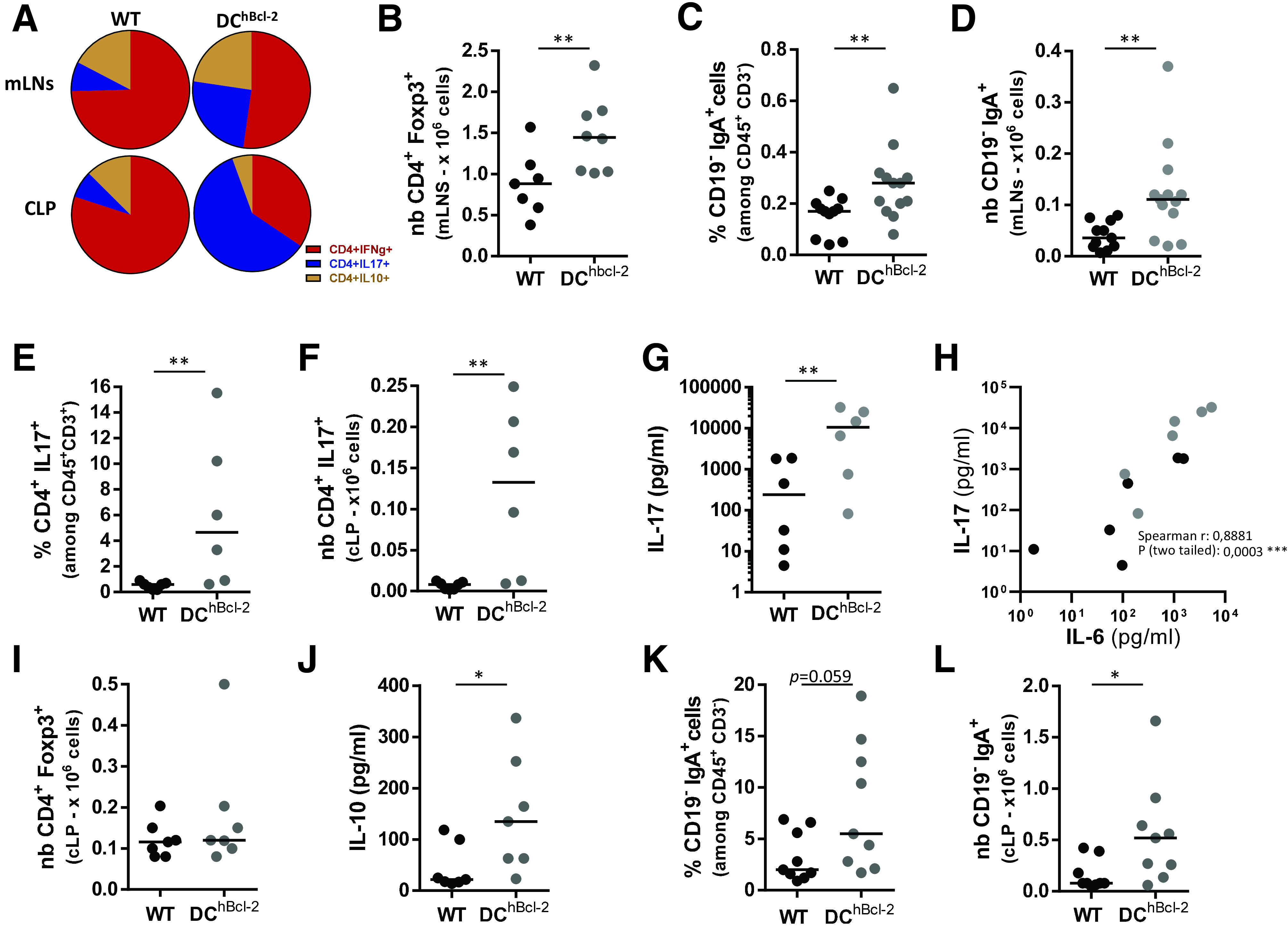
Figure 4.

HFD-fed DChBcl-2 mice showed enhanced Treg, Th17, and sIgA+ B-cells that established in the colon. All data are representative of mice fed an HFD for 24 weeks. A: Circle graphs representing the mean proportions of interferon-γ (IFN-γ–), IL-17–, and IL-10–producing CD4+ T cells in the mLNs or CLP after intracellular staining of cytokines. Total numbers (nb) of CD4+Foxp3+ T lymphocytes in the mLNs (B) or in the CLP (I). Proportions of CD19−IgA+ plasmablasts in the mLNs (C) or in the CLP (K). Total numbers of CD19−IgA+ plasmablasts in the mLNs (D) or the CLP (L). Percentages (E) and total numbers (F) of IL-17–producing CD4+ T cells in the CLP after intracellular staining of cytokines. IL-17 (G) and IL-10 (J) secretion in the supernatants of ex vivo anti-CD3/CD28–stimulated CLP cells for 72 h. H: Correlation graph of IL-17 and IL-6 cytokines secreted in the supernatants of ex vivo anti-CD3/CD28–stimulated CLP cells for 72 h. Data are presented as mean for circle graphs or median for dot plots. *P < 0.05; **P < 0.01.
