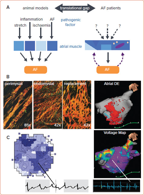
Figure 1: Obstacles to Translation of Results from Large-animal Models of AF.

A: In animal models, pathogenic stimuli are applied at high intensity for a relatively short duration to young healthy animals. AF patients are often elderly and have an extensive history of underlying risk factors for AF. B: In animal studies, the availability of tissue samples allows detailed characterisation of tissue properties, including the distribution of various types of fibrosis. In patients, delayed enhancement imaging of the thin atrial wall has been used to visualise the distribution of fibrosis as a binary parameter (scar versus normal myocardium).[11,33] C: In open-chest animal studies, high-density epicardial mapping of unipolar electrograms has allowed detailed reconstruction of complex fibrillation patterns, whereas (sequential) voltage mapping using bipolar electrograms in patients offers a less detailed view.[33,95] DE = delayed enhancement. Sources: B: Adapted from Weber et al. 1989.[11] Used with permission from Elsevier. B and C: Adapted from Chen et al. 2019.[33] Used with permission from Oxford University Press. C: Adapted from Verheule et al. 2010.[95] Used with permission from Wolters Kluwer Health.
