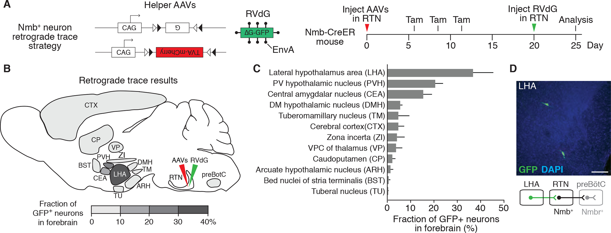
Figure 2. Mapping the direct forebrain inputs to RTN Nmb neurons by monosynaptic retrograde tracing.

(A) Scheme for rabies virus monosynaptic retrograde tracing of inputs to Nmb neurons. Two Cre-dependent helper AAVs (CAG-FLExloxP-rabies glycoprotein, top, and CAG-FLExloxP-TVA:mCherry, bottom) were injected into RTN (see B) of Nmb-CreER mouse, followed by intraperitoneal tamoxifen (Tam) injections to express in Nmb-CreERT2-expressing neurons the rabies glycoprotein (G) and the TVA receptor for EnvA fused to mCherry. After injection of EnvA-pseudotyped, glycoprotein deleted and GFP-expressing rabies virus (RVdG) to the same region (RTN), direct inputs to Nmb neurons are labeled. Open and closed triangles, Cre recombination sites. (B and C) Quantification of forebrain regions labeled by monosynaptic retrograde tracing in A that provide direct input to RTN Nmb neurons. Locations of labeled regions are density-coded in B to show relative abundance of labeling. DMH, dorsomedial hypothalamic nucleus; PVH, paraventricular hypothalamic nucleus; VPC, ventral posterior complex. Mean +/−SEM, n=3. (D) Coronal section (30 um) of mouse brain showing two rabies-labeled GFP-positive neurons in LHA region and a schematic below of their projection (green) from LHA to RTN. preBötC, preBötzinger Complex. Scale bar, 100 um.
