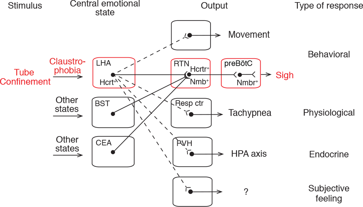
Figure 7. Model of the circuit organization of claustrophobia and its relation to other emotional circuits.

Tube confinement activates HCRT neurons in LHA, which directly activate Hcrtr+ Nmb+ double-positive neurons in RTN, which in turn project to and directly activate Nmbr+ neurons in the preBötC, comprising a neuropeptide “relay circuit” that triggers sighing. The LHA HCRT neurons represent a key node in the central emotional state of claustrophobic stress because they also activate other behavioral (struggling movements), physiological (tachypnea), and endocrine (hypothalamic-pituitary-adrenal axis, HPA axis) outputs, and perhaps subjective feelings through other targets (dashed lines). Other central emotional states (e.g., relief, sadness) are associated with activation of other brain regions, some of which (e.g., bed nuclei of stria terminalis, BST; central amygdalar nucleus, CEA) also project to the Nmb+ RTN neurons (solid lines) and induce sighing. Certain physiological states (e.g., hypoxia) also provide input to the Nmb+ RTN neurons (not shown). Resp ctr, respiratory center (not RTN); PVH, paraventricular hypothalamic nucleus.
