Figure 1.
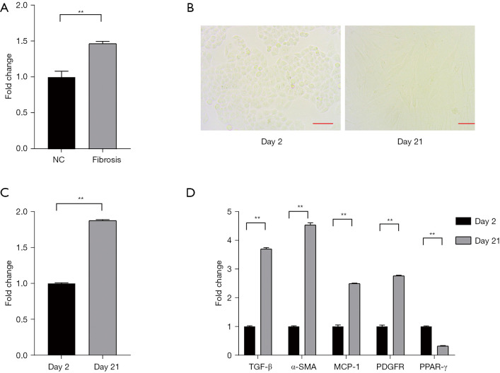
MiR-34a expression was increased in liver fibrosis tissue. (A) The level of miR-34a in liver tissue; (B) the fibrosis morphology of HSCs cultured in vitro changed over time; (C) miR-34a was up-regulated in the in vitro cultured HSCs at 21 days; (D) TGF-β, α-SMA, MCP-1, and PDGFR were up-regulated on day 21. Scale bar =100 µm. **, P<0.01. HSCs, hepatic stellate cells.

