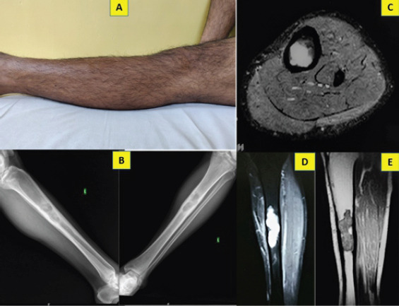
Figure 1.

Pre-operative clinical picture of the left leg with no appreciable swelling or skin changes (a); anteroposterior and lateral plain radiographs showing multiloculated, lytic lesion in the diaphysis with cortical thickening and widening of medullary cavity (b); STIR (c, d) and T1-weighted (e) MRI images showing focal area of lobular intramedullary lesion, endosteal scalloping and cortical thickening without extra osseous extension.
