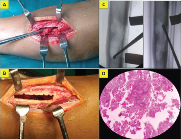
Figure 2.

Intraoperative picture showing extended curettage of the lesion (a, b, c) through a cortical window and the pathology slide showing hypo cellularity and lacunar spaces harboring binucleate chondrocytes with intervening chondroid matrix (d).
