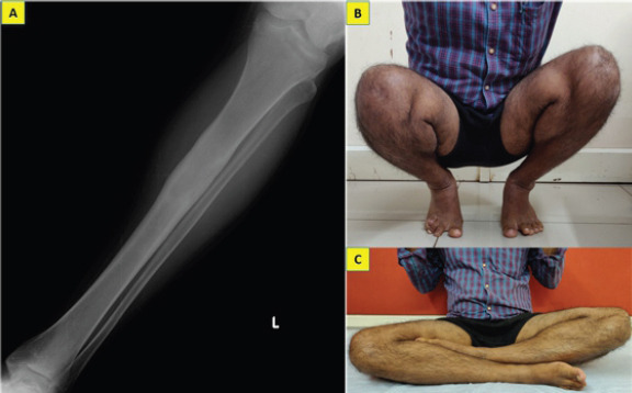
Figure 3.

Anteroposterior radiographs of the left tibia taken at 2-year follow-up showing complete healing of the lesion without evidence of recurrence (a) and clinical photograph demonstrating his ability to squat and sit cross-legged normally which are essential in his occupation (b, c).
