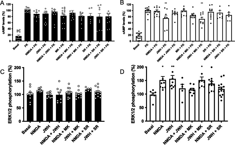
Fig. 4.
Signaling in primary microglia activated with LPS and IFN-γ. Primary microglial cells were incubated for 48 h in the absence (black bars) or in the presence (white bars) of 1 μM LPS and 200 U/mL IFN-γ. Cells were treated with selective agonists (15 μM NMDA for NMDA channel, and/or 100 nM JWH-133 for CB2R) and cAMP levels and MAPK pathway activation were determined. As Gi coupling was assessed, decreases in cAMP levels were determined in cells previously treated with 0.5 μM forskolin (15 min). When indicated cells were pretreated with selective receptor antagonists (1 μM MK-801 for NMDA or 1 μM SR-144528 for CB2R). Values are the mean ± S.E.M. of 5 independent experiments performed in triplicates. One-way ANOVA followed by Bonferroni’s multiple comparison post hoc test were used for statistical analysis (*p < 0.05, **p < 0.01, ***p < 0.001 versus forskolin treatment in cAMP determinations or versus vehicle treatment (basal) in ERK phosphorylation assays). ANOVA summary: A F: 12.0, p<0.001; B F: 30.0, p<0.001, C F: 1.8, p<0.093, D F: 4.1, p<0.001

