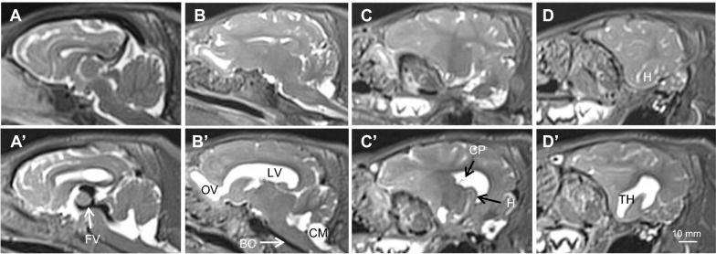
Fig. 1.
Representative T2-weighted sagittal MRI images summarizing brain and ventricular morphology in non-hydrocephalic control (A–D) and hydrocephalic kaolin-injected pre-shunt conditions (A′–D′); panels arranged left to right from midsagittal (A, A′) to lateral (D, D′). The intact control piglet (case 25) is 41-days old. Pre-shunt images are taken from case 13 at 18-days post-kaolin. In the pre-shunt condition, note the kaolin blockage of the basal cisterns (BC), prominent flow void (FV, black) within the third ventricle and cerebral aqueduct indicative of high CSF pulsatility, the patent channel connecting the olfactory ventricle (OV) to the lateral ventricle (LV), the choroid plexus (CP) floating in the LV, and the enlargement of all cerebral ventricles, especially the temporal horns (TH) containing the hippocampus (H), but no periventricular edema. The cisterna magna (CM) remains open. Scale bar = 10 mm for each all panels

