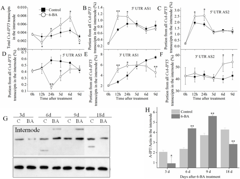
Fig. 3.
Expression of CsA-IPT5 transcripts and CsA-IPT5 protein in the internode induced by 6-BA application. A Expression of total CsA-IPT5 transcripts in the internode at different time points induced by 6-BA. B-F The ratios of CsA-IPT5 splice variants in the internode at different time points induced by 6-BA. Western blot (G) and ‘CsA-IPT5/Actin’ (H) which showed CsA-IPT5 protein expression in the internode at 3 d, 6 d, 9 d and 18 d after 6-BA treatment. Data are means ±SD (n = 3 or 6). For figures A-F, asterisks indicate significant differences in each index between control and 6-BA treatment at each time point; for figure H, asterisks indicate significant differences in A-IPT/Actin between control and 6-BA treatment at each time point (**P < 0.01; Student’s t-test)

