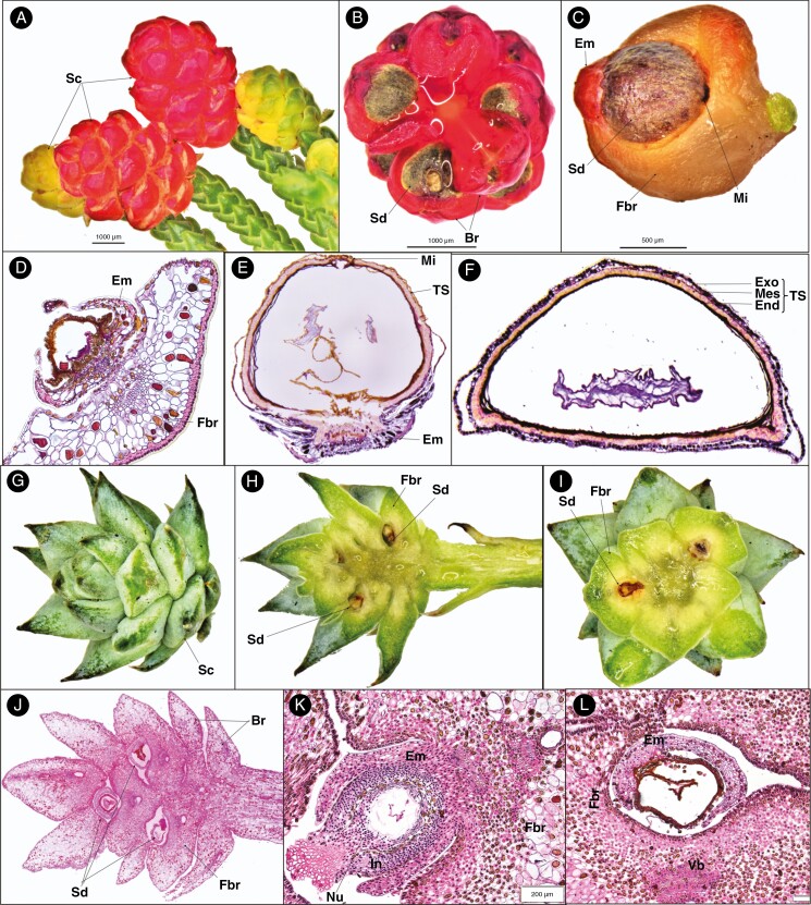
Fig. 3.
Seed cone morpho-anatomical features of Microcachrys (A–F) and Saxegothaea (G–L) showing: (A) fleshy red and young yellowish seed cones; (B) seed cone cross-section view showing seed (Sd) and bract (Br); (C) seed cone with epimatium (Em), seed (Sd), fertile bract (Fbr) and micropyle (Mi); (D) young ovule development (post-pollination) shows fertile bract, epimatium and ovule (Ov); (E) longitudinal section of single seed cone showing testa (TS), micropyle (Mi) and epimatium; (F) seed cone cross-section showing the different layer of testa, i.e. exotesta (Exo), mesotesta (Mes) and endotesta (End); (G) seed cone (Sc); (H) seed cone longitudinal section showing fertile bract (Fbr) and seed (Sd); (I) cross-sections showing the seed cone with the fertile bract and seed; (J) longitudinal section of seed cone showing fertile bract, seed and bract; (K) longitudinal section of developing ovule (post-pollination) showing epimatium, nucellus (Nu), fertile bract and integument (In); and (L) cross-section of developing ovule (post-pollination) showing epimatium, fertile bract and vascular bundle.

