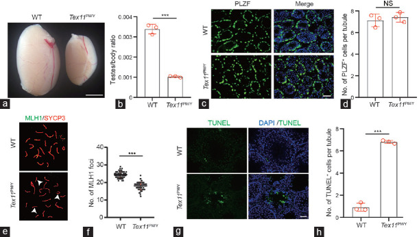
Figure 1.
Male mice carrying an azoospermia patient-derived TEX11 mutation display meiotic arrest. (a) Testis morphology of WT and Tex11PM/Y mice. Scale bar = 100 mm. (b) Relative testis/body weight of WT and Tex11PM/Y mice. The average values of three separate experiments are plotted. Error bar: s.d. Significance was determined by two-tailed, unpaired Student’s t-test. (c) Immunostaining of 10 dpp testis sections with an anti-PLZF antibody. Green: PLZF; Blue: Hoechst 33342. Scale bar = 50 μm. (d) Quantification of PLZF+ cells in 10 dpp testis sections. A total of 237 and 222 tubules from three WT and Tex11PM/Y mice were scored. Error bar: s.d. Significance was determined by two-tailed, unpaired Student’s t-test. NS: no significance. (e) Immunostaining of MLH1/SCP3 in spermatocyte spreads prepared from 8-week-old WT and Tex11PM/Y mice. Green: MLH1; red: SYCP3. Note the absence of MLH1 foci on three synapsed chromosomes (arrowhead) in the Tex11PM/Y sample shown in the bottom image. (f) Number of MLH1 foci in Tex11PM/Y and WT spermatogonia. A dramatic reduction in the number of MLH1 foci (green) in Tex11PM/Y spermatogonia (approximately 15 foci per cell) relative to WT (22 foci per cell). Error bar: s.d. Significance was determined by two-tailed, unpaired Student’s t-test;*** P < 0.001. (g) TUNEL analysis of Tex11PM/Y and WT testis. Massive apoptosis (green) shown in Tex11PM/Y tubules. Scale bar = 100 μm. (h) Analysis of apoptotic cells in Tex11PM/Y and WT testis. The average values of three separate experiments are plotted. Error bar: s.d. Significance was determined by two-tailed, unpaired Student’s t-test;*** P < 0.001. TEX11: testis-expressed 11; WT: wild-type; PLZF: promyelocytic leukemia zinc finger; MLH1: mutl homolog 1; SYCP3: synaptonemal complex protein 3; TUNEL: terminal deoxynucleotidyl transferase biotin-dUTP nick end labeling; DAPI: 4’,6-diamidino-2-phenylindole; s.d.: standard deviation; dpp: days postpartum.

