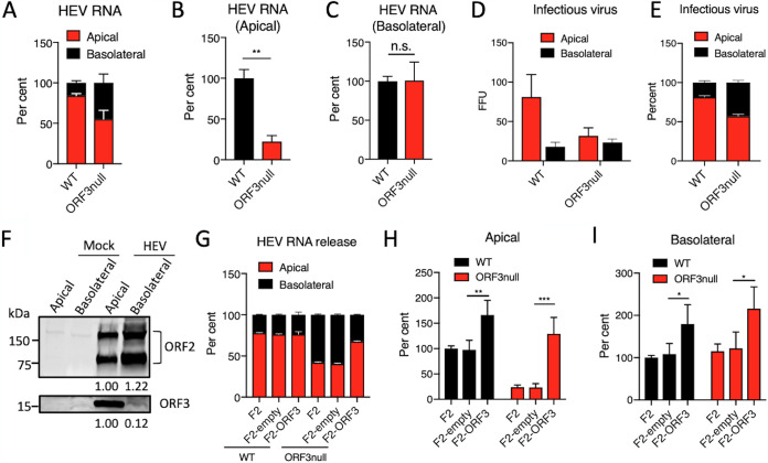
FIG 3.
ORF3 facilitates apical release of HEV particles from polarized human hepatocytes. (A to C) Polarized F2 cells were inoculated with WT or ORF3null HEV (5 × 10³ GE per cell). After 7 days, HEV RNA in the culture supernatants from the top (apical) and lower (basolateral) chambers and intracellular HEV RNA were isolated and quantified by qRT-PCR. (A) Relative amounts of HEV RNA present in the supernatants from the apical versus basolateral chambers. (B and C) Relative amounts of WT versus ORF3null HEV RNA present in the apical (B) and basolateral (C) chambers (extracellular HEV RNA levels were normalized to intracellular HEV RNA levels to adjust the difference in virus replication between WT and ORF3null HEV, and the values from the WT HEV-infected group were set as 100%). Data are shown as means ± SEM from at least 2 independent experiments, each with duplicate wells. n.s., not significant; **, P < 0.01. (D and E) Polarized F2 cells were inoculated with WT or ORF3null HEV (5 × 10³ GE per cell), and culture supernatants in the apical and basolateral chambers were collected at 5 dpi to inoculate naive F2 cells. (D) Cells were fixed 5 days later, and ORF2-positive focus-forming units (FFU) were determined by immunofluorescence assays. (E) Relative amounts of infectious virus present in the apical versus basolateral compartments. Data are shown as means ± SEM from triplicate samples. (F) Western blots of ORF2 and ORF3 proteins in culture supernatants from the apical and basolateral chambers of WT HEV (5 × 10³ GE per cell)-infected polarized F2 cell cultures. Note that the secreted ORF2 protein existed in both monomeric and dimeric forms. Values below each blot indicate the relative ORF2 or ORF3 protein levels. (G to I) Polarized F2, F2-empty, and F2-ORF3 cells were inoculated with WT or ORF3null HEV (5 × 10³ GE per cell). After 7 days, HEV RNAs in culture supernatants (in both the apical and basolateral chambers) and cells were isolated and quantified by qRT-PCR. (G) Relative amounts of HEV RNA present in the supernatants from the apical versus the basolateral chambers. (H and I) Relative amounts of WT versus ORF3null HEV RNA released in the apical (H) and basolateral (I) chambers (values from WT-infected F2 cells were set as 100%). Data are shown as means ± SEM from 2 independent experiments, each with duplicate wells. *, P < 0.05; **, P < 0.01; ***, P < 0.001.

