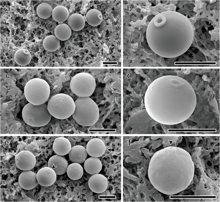
FIGURE 5.
Scanning electron micrographs of three new Spumella species. (a,b) SEM images of the spherical stomatocyst of S. longicolla Yangrim041319B6. (c,d) SEM images of the oblate stomatocyst of S. oblata Sanggul120118B3. (e,f) SEM images of the spherical stomatocyst of S. rotundata Woeam020118A5. Arrowheads indicate a pore of the stomatocyst. Scale bars = 5 μm.

