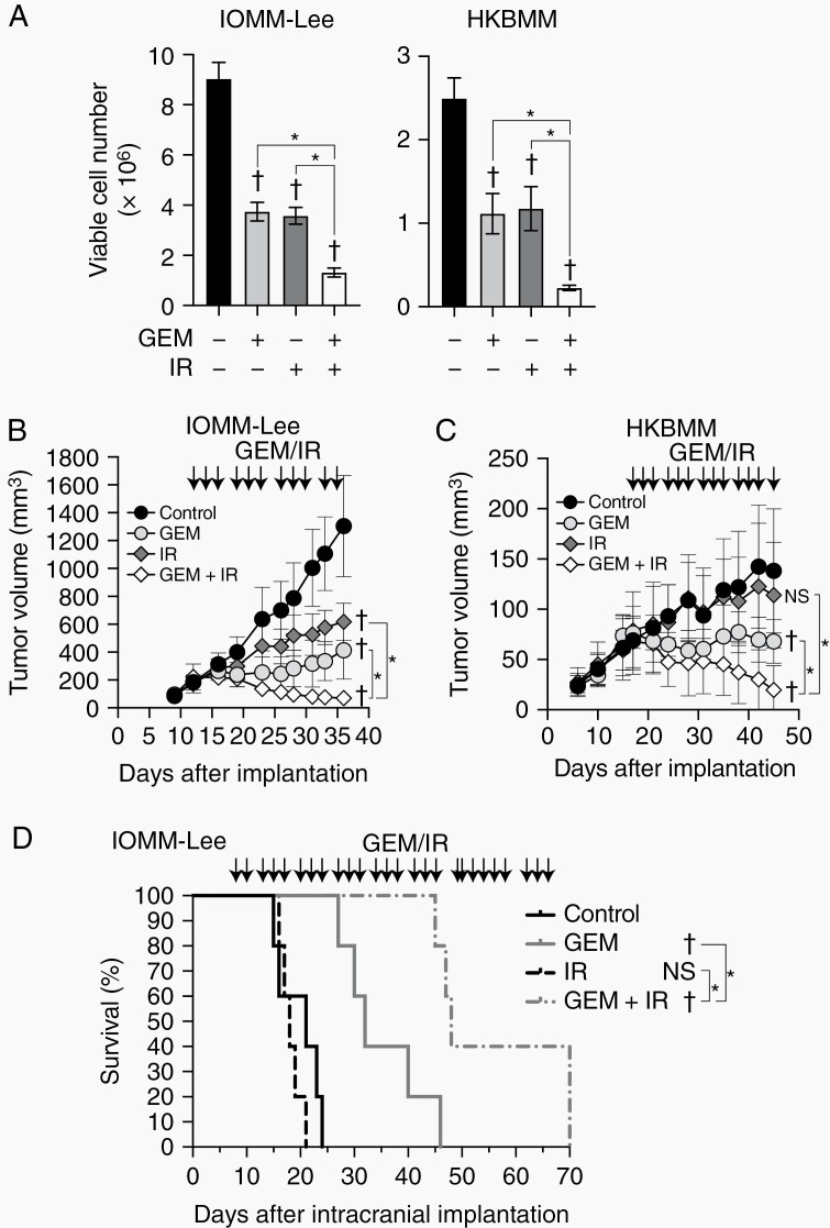
Figure 1.
Combined effects of gemcitabine with ionizing radiation on malignant meningioma cells in vitro and in vivo.
(A) IOMM-Lee and HKBMM cells plated on 6-well plates in 6 replicates (2 × 10⁵ cells per well for IOMM-Lee and 4 × 10⁵ cells per well for HKBMM) were incubated without or with gemcitabine (3 nM for IOMM-Lee and 2 nM for HKBMM) for 6 days and not irradiated or irradiated 3 times by X-ray (1 Gy for IOMM-Lee and 2 Gy for HKBMM) on days 1, 3, and 5, and subjected to a cell viability assay. (B and C) IOMM-Lee and HKBMM cells were subcutaneously implanted in the flank regions bilaterally (1 × 10⁶ for IOMM-Lee and 2 × 10⁶ for HKBMM). After tumor establishment was confirmed, mice were treated with gemcitabine (10 mg/kg, intraperitoneal injection) (GEM), ionizing radiation (1 Gy) (IR), both (GEM+IR), or vehicle (Control) 3 times a week (arrows). The size of tumors was measured (n = 8 for each group). (D) Eight days after the intracranial implantation of IOMM-Lee cells (2 × 10⁵ cells per mouse), mice were treated with gemcitabine (10 mg/kg) (GEM), ionizing radiation (1 Gy) (IR), both (GEM + IR), or vehicle (Control) 3 times a week (arrows). The survival of mice was shown as the Kaplan-Meier plot (n = 5, each group). GEM, gemcitabine. IR, ionizing radiation. Values are shown as mean ± SD. In (A), (B), and (C), P-values were calculated by a 1-way ANOVA with Tukey’s post hoc test. In (D), P-values were calculated by the Log-rank test. *P < .05. †P < .05 and NS, P ≥ .05 vs the Control (GEM− and IR−).

