Figure 5.
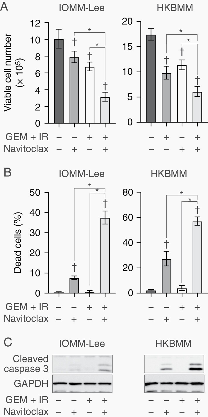
Effects of navitoclax combined with gemcitabine and ionizing radiation on malignant meningioma cells.
IOMM-Lee and HKBMM cells plated on 6-well plates in 6 replicates (2 × 10⁵ cells per well for IOMM-Lee and 4 × 10⁵ cells per well for HKBMM) were incubated for 2 days without or with gemcitabine (3 nM for IOMM-Lee and 2 nM for HKBMM) in combination with ionizing radiation (1 Gy for IOMM-Lee and 2 Gy for HKBMM) on day 1 in the absence or presence of navitoclax (1 µM) and were then subjected to a cell viability assay to assess the viable cell number (A) and percentage of dead cells (B) or to a Western blot analysis to examine the expression of indicated proteins (C) on day 2. Values are shown as mean ± SD. P-values were calculated by a 1-way ANOVA with Tukey’s post hoc test. *P < .05. †P < .05 vs the Control (GEM + IR− and Navitoclax−).

