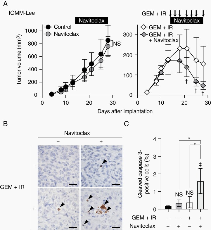
Figure 6.
Effects of navitoclax on malignant meningioma xenografts in combination with gemcitabine and ionizing radiation.
(A) IOMM-Lee cells were subcutaneously implanted in the flank regions bilaterally (1 × 10⁶). After tumor establishment was confirmed, mice were treated with navitoclax (100 mg/kg, oral gavage, every day), gemcitabine (10 mg/kg, intraperitoneal injection) in combination with ionizing radiation (1 Gy) (3 times a week, arrows) (GEM + IR), both (GEM + IR + Navitoclax), or vehicle (Control). The size of tumors was measured. Values are shown as mean ± SD (n = 8 for each group). (B) Immunohistochemistry for cleaved caspase 3 in tumors excised 1 day after the last treatment. Arrowheads indicate cleaved caspase 3-positive cells. Scale bars, 50 µm. (C) Quantification of the percentages of cleaved caspase 3-positive cells. In (A), P-values were calculated by a 2-way ANOVA with the Bonferroni multiple comparisons test. NS, P ≥ .05 and †P < .05 vs GEM + IR. In (C), P-values were calculated by a 1-way ANOVA with Tukey’s post hoc test. *P < .05. ‡P < .05 and NS, P ≥ .05 vs the Control (no treatment).

