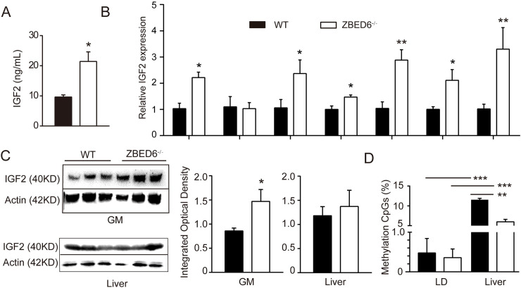
Fig 3. Increased levels of IGF2 in multiple tissues except liver in WT and ZBED6-/- pigs (n = 3).
Gastrocnemius muscle (GM), longissimus dorsi (LD). (A) Serum concentrations of IGF2 in WT and ZBED6-/- pigs at 3 months (B) qPCR analysis of IGF2 mRNA in multiple tissues from ZBED6-/- pigs. (C) Western blot analysis of IGF2 in GM and liver of ZBED6-/- pigs. Results of western blot were quantified by Iamge J. The relative levels of IGF2 protein in GM and liver were plotted. (D) Percentage methylation around IGF2-intron 3–3072 (GCTCG) of LD and liver in WT and ZBED6-/-. The results are the means ± SEMs. *p < 0.05; **p < 0.01, *** p < 0.001, Student’s t test.

