. 2021 Nov 9;10:e71683. doi: 10.7554/eLife.71683

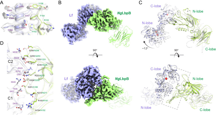
Figure 4. The 3.65 Å cryoEM structure of N. gonorrhoeae LbpB (NgLbpB) in complex with human lactoferrin.

(A) Zoomed view at the interface between NgLbpB and lactoferrin (Lf) depicting the quality of the density shown as a grey isosurface. (B) Orthogonal views of the full cryoEM map with NgLbpB in green and Lf in violet. (C) Orthogonal view of an alignment of the NgLbpB–Lf cryoEM structure (green/violet) with the N. meningitidis LbpB (NmLbpB)–Lf crystal structure (grey) (RMSD 1.4 Å along the interacting domains). (D) A zoomed view of the binding interface shows extensive interactions along an elongated surface covering both the C1 and C2 domains of Lf (buried surface area 1604 Å2).

Figure 4.

Figure 4—figure supplement 1. CryoEM data processing workflow for N. gonorrhoeae LbpB (NgLbpB)–Lf complex.
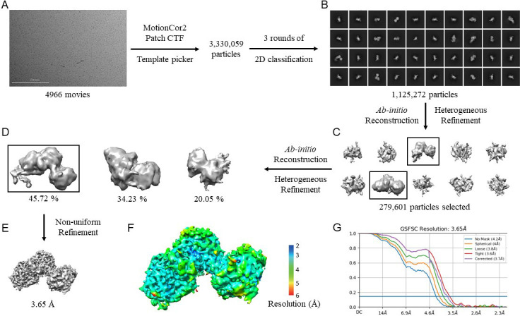
Figure 4—figure supplement 1.

(A) Representative cryoEM micrograph from a total of 4966. Picked particles were subjected to iterative rounds of 2D classification. (B) Representative 2D class averages show different orientations of the particles. Two rounds of ab initio reconstruction followed by heterogeneous refinement produced 10 initial models in round 1 (C) and 3 initial models in rounds 2 (D). Boxed classes were selected for further processing. (E) Final 3D reconstruction map upon non-uniform refinement was obtained at 3.65 Å. (F) Local resolution of final cryoEM map. (G) Gold-standard Fourier shell correlation (GSFSC) curve of NgLbpB–Lf map. The horizontal blue line indicates 0.143 cutoff for resolution estimation.
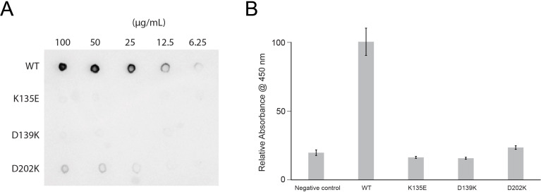
Figure 4—figure supplement 2. Lactoferrin binding to N. gonorrhoeae LbpB (NgLbpB) and NgLbpB mutants.

Figure 4—figure supplement 2.

(A) Solid-phase-binding assays of dilutions of NgLbpB and mutants with horse radish peroxidase (HRP)-conjugated lactoferrin probe. (B) Enzyme-linked immunosorbent assays (ELISAs) of NgLbpB and mutants showing normalized absorbance. All experiments were done at least in triplicate.
Figure 4—figure supplement 2—source data 1. Lactoferrin binding to N. gonorrhoeae LbpB (NgLbpB) and NgLbpB mutants.
(A) Solid-phase-binding assays ofdilutions of NgLbpB and mutants with horse radish peroxidase (HRP)-conjugated lactoferrin probe. (The red arrow indicates the original blot that was cropped for this panel.) (B) Enzyme-linked immunosorbent assays (ELISAs) of NgLbpB andmutants showing normalized absorbance. All experiments were done at least in triplicate.
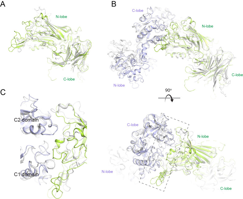
Figure 4—figure supplement 3. Structural comparison of N. meningitidis LbpB (NmLbpB) to NmTbpB.

Figure 4—figure supplement 3.

(A) A superposition of the NmLbpB (green) and NmTbpB (grey) (PDB ID 3V8U) structures (RMSD of 2.0 Å). (B) Orthogonal views of a superposition of the NmLbpB–lactoferrin (Lf) (green/violet) and NmTbpB–transferrin (Tf) (grey) (PDB ID 3VE1) structures (overall RMSD of 4.0 Å; an RMSD of 1.4 Å was calculated for alignment of the interacting domains only). (C) A zoomed view from panel B of the dashed box along the binding interfaces.