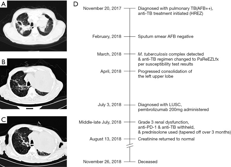
Figure 2.
Representative chest CT images at different stages of disease course (A-C) and timeline of therapy and disease status for both lung cancer and TB infection in patient P1 (D). (A) shows patchy shadows in both lungs (including cavity in the right lung), tracheobronchial stenosis in the left upper lobe, and soft tissue shadows. Chest CT images before the initiation of anti-PD-1 immunotherapy (B, TB lesions on the right were absorbed remarkably, but the consolidation of the left upper lobe progressed) and 2 months after the initiation of immunotherapy (C). TB, tuberculosis; AFB, acid-fast bacillus; HREZ, isoniazid, rifampicin, ethambutol, and pyrazinamide; PaReEZLfx, pasiniazid, rifapentine, ethambutol, pyrazinamide, and levofloxacin; LUSC, lung squamous cell carcinoma; PD-1, programmed cell death 1.

