FIG. 4.
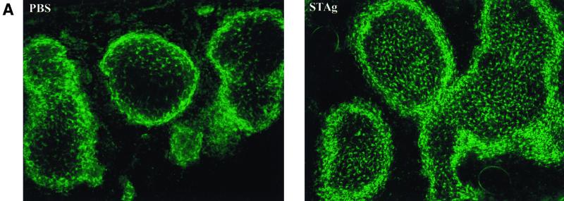
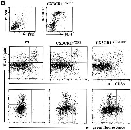
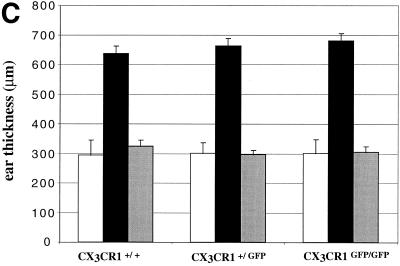
Analysis of CX3CR1 function in DC. (A) Cryosection of paraformaldehyde-fixed spleens of CX3CR1GFP/GFP mice 6 h after PBS or STAg injection indicating STAg-induced recruitment of DC to central periarteriolar lymphoid sheaths. Note that there is no depletion of GFP-positive cells from the marginal zone in the STAg-injected spleen due to recruitment of CD11b+ CD11c− blood monocytes. (B) Flow cytometric analysis of overnight-cultured DC isolated from STAg-injected spleens of wt, CX3CR1+/GFP, and CX3CR1GFP/GFP mice. Cells are gated according to scatter and CD11c expression as indicated. The fractions of IL-12-producing CD8+ DC were 50%, 50% (± 2.4%), and 49% (± 12%) for wt, heterozygous, and mutant mice, respectively. Note the absence of IL-12 (p40)-positive cells among the GFPbright DC of CX3CR1+/GFP and CX3CR1GFP/GFP mice, indicating that the FKN-positive CD8+ DC do not participate in IL-12 production. (C) Contact hypersensitivity assay. Data represent mean (± standard deviation) of results obtained from age-matched wt BALB/c mice and CX3CR1+/GFP and CX3CR1GFP/GFP BALB/c mice (N6) (n = 5 per time point). Open bars, ear thickness before challenge (day 6); black bars, ear thickness 24 h after oxazolone challenge; grey bars, control ear thickness 24 h after challenge with vehicle only.


