FIG. 5.
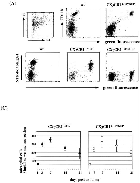
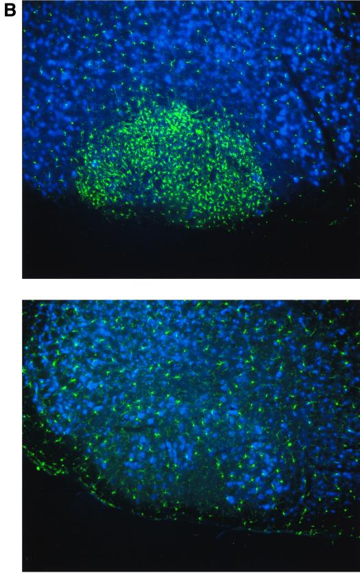
Analysis of CX3CR1 function in microglia. (A) Surface CX3CR1 staining of microglial cells isolated via Percoll density gradient from collagenase-digested brains of wt, CX3CR1+/GFP, and CX3CR1GFP/GFP mice. Cells were stained for the indicated surface markers (CD11b and CX3CR1) and gated according to scatter and viability as indicated. (B) Peripheral nerve transection experiment. Coronal section through operated and contralateral control facial nerve nuclei of axotomized CX3CR1+/GFP mouse day 7 after axotomy. Section were stained with an anti-neuronal nucleus-specific antibody (NeuN) and Cy5-coupled sheep anti-mouse IgG serum. (C) Quantitative evaluation of microglial reaction in response to facial nerve transection in operated CX3CR1+/GFP and CX3CR1GFP/GFP mice. The volume analyzed in the facial nerve nucleus cross sections represents about 0.25 mm2 by 12 μm. Results are presented as means (± standard deviations) of 16 sections obtained from four mice of each genotype per time point.


