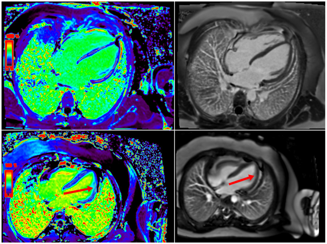
Figure 1.

Extracellular volume (ECV) map and late gadolinium enhancement (LGE) in patients without systolic dysfunction. (Top) ECV map and LGE image showing normal ECV and no LGE. (Bottom) ECV map showing increased ECV signal in one region of interest (red arrow) without any evidence of LGE (red arrow on right).
