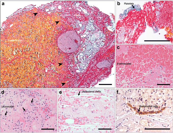
Fig. 1.
Coronary atherothrombotic specimens from patients undergoing treatment for ST-segment elevation myocardial infarction. The border zone between cholesterol cleft rich atheroma and thrombus is evident (arrows) with Carstairs staining rendering erythrocytes, yellow, and fibrin, pink (a). Atherothrombotic specimens were composed of platelets (blue) (b), erythrocytes (c), leucocytes (d), cholesterol clefts (e), and CD146+ endothelial cells (f) encased in fibrin. Scale bars 50 μm

