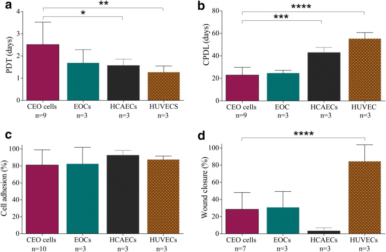
Fig. 3.
Proliferation, adhesion, and wound migration of coronary endothelial outgrowth cells in vitro. Coronary endothelial outgrowth (CEO) cells had a higher population doubling time (PDT) during the first 9 passage in comparison to human coronary artery endothelial cells (HCAECs) and human umbilical vein endothelial cells (HUVECs) (one-way analysis of variance P = 0.017; Bonferroni post-test *P < 0.05 and **P < 0.01, respectively. n = 3–9) (a) and a lower cumulative population doubling level (CPDL) when maintained long term in culture (P < 0.0001; Bonferroni post-test ***P < 0.001 and ****P < 0.0001, respectively. n = 3–9) (b). CEO cells had a similar capacity to adhere to a collagen substrate in vitro (P = 0.662. n = 3–10) (c), but following the infliction of a linear wound in the endothelial cell monolayer, CEO cells had a reduced capacity to migrate in vitro in comparison to HUVECs (P = 0.008; Bonferroni post-test ****P < 0.0001. n = 3–7) (d)

