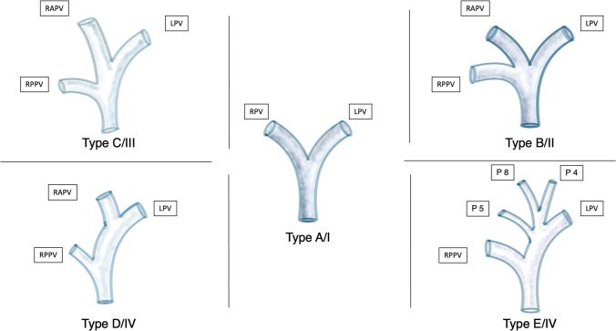
Fig. 2.
Classification of the portal vein (PV) according to the classifications proposed by Nakamura et al. [32] (Type A to E) and Cheng et al. [33] (Type I to IV). Type A/I Normal anatomy (common variant): bifurcation of the main portal vein (PV) into the left portal vein (LPV) and right portal vein (RPV) (80%) [34]. Type B/II Anatomic variation of a trifurcation of the main PV into the LPV, the right anterior portal vein (RAPV), and the right posterior portal vein (RPPV). A common RPV is missing (7–16%) [8]. Type C/III RPPV arises separately from the main portal vein followed by extraparenchymal bifurcation into the RAPV and LPV intraparenchymal (5%). Type D/IV: RPPV arises separately from the main portal vein followed by intraparenchymal branching of the RAPV. Type E/IV: Separate portal vein branches for liver segments 4, 5, and 8

