
Figure 3. Vaginal Configuration and Displacement in the Anatomic Space.
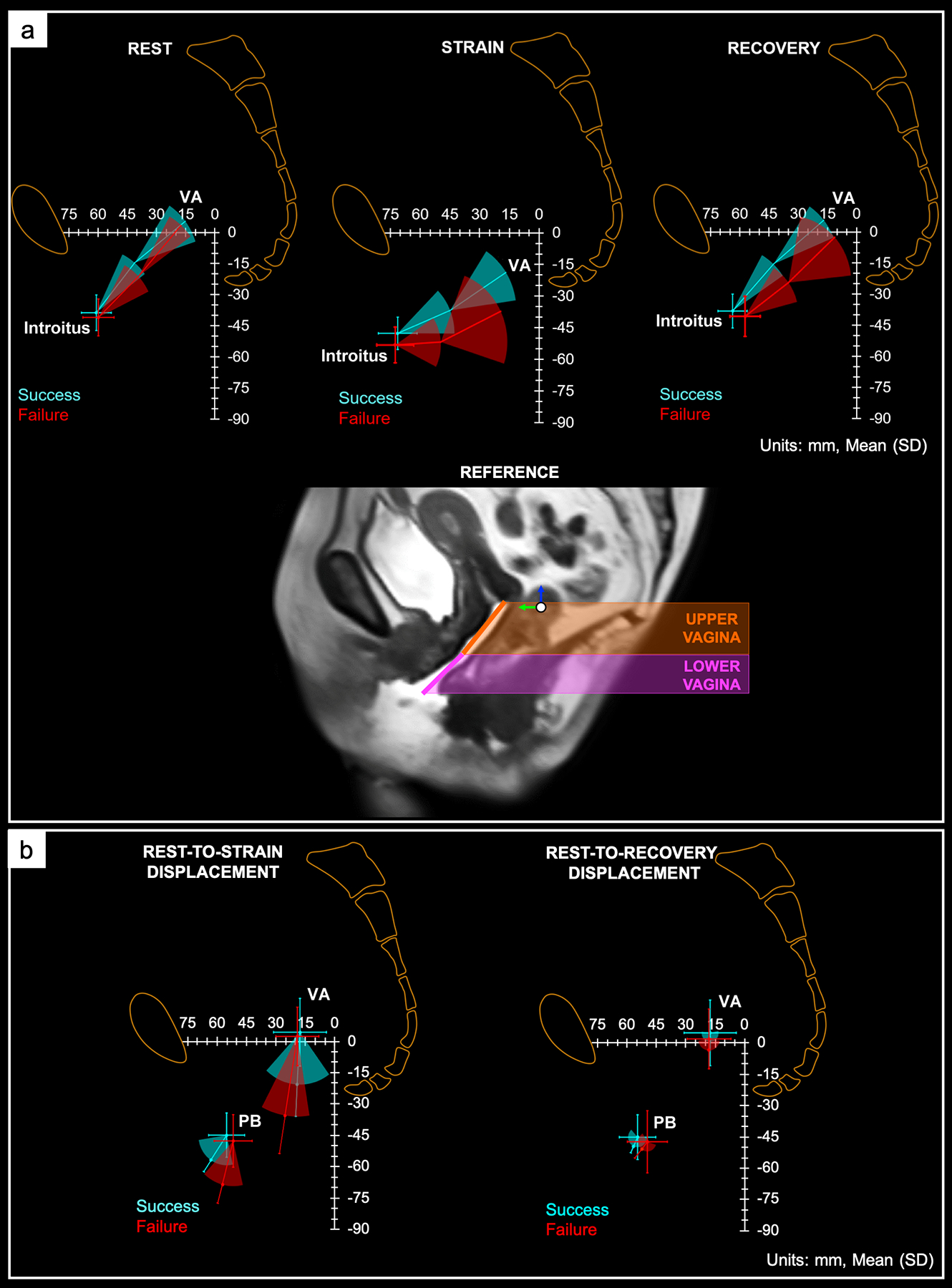
Visual representation of the change in orientation and position of the upper and lower vagina with respect to the pelvic space at rest (left), maximal strain (middle), and recovery (right) in magnetic resonance imaging (MRI) successes and failures. The orientation (i.e., angulation) of the upper and lower vagina and the position of the vaginal apex (VA), and mid-point of the vaginal introitus (introitus) are shown. More posterior-inferior deviation of the upper and lower vagina was observed in MRI failures, signifying poorer apical, mid, and distal vaginal support compared to successes. Data are presented as mean and standard deviation. Units are in mm. (b) Position and displacement vector (mean and standard deviation) of the vaginal apex (VA) and perineal body (PB) from rest to strain (left) and rest to recovery (right). MRI successes (blue) and failures (red) are indicated. The magnitude of the VA and PB displacement from rest to strain was greater in MRI failures vs successes, suggesting less apical and distal vaginal support in failures compared to successes. Rest-to-recovery VA and PB displacement was similar in both outcome groups. Units are in mm.