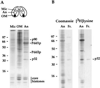
FIG. 1.
Polypeptides synthesized and targeted to different nuclei during late stages of Tetrahymena development. (A) Conjugating cells (schematically represented on the top of the figure) were pulse-labeled with [3H]lysine from 9 to 10 h postmixing. Nuclei were purified by sedimentation at 1 U of gravity (2). Total protein from different types of nuclei was resolved on a 12% SDS–polyacrylamide gel and analyzed by fluorography. Approximately 107 micronuclei (Mic), 106 old macronuclei (OM), and 3 × 10⁶ anlagen (An) were used. Previously identified Tetrahymena polypeptides and protein molecular mass standards (kilodaltons) are shown. (B) p32 recovered from reverse-phase HPLC fractionation of proteins extracted from purified anlagen and resolved on 12% SDS–polyacrylamide gel. Proteins were visualized by staining with Coomassie blue, followed by fluorography to verify the position of p32 (shown on the right). An, total anlagen protein before the extraction; Fr, HPLC fraction containing p32. Protein molecular mass standards (kilodaltons) are designated on the left.

