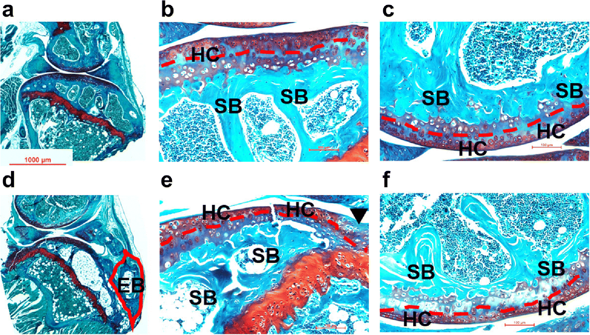
Fig. 2. Safranin O and Fast Green staining of mouse knee joints at week 16 after surgery.

Microphotographs of sham (a: whole joint; b: tibia; c: femur) and ACLT (d: whole joint; e: tibia; f: femur) samples were taken under a microscope. Compared to the sham joints, various degenerative changes were found in ACLT joints, including reduced hyaline cartilage (red dash line), erosion of articular cartilage (black arrow head) and formation of ectopic bone (red line frame). EB: ectopic bone; HC: hyaline cartilage; SB: subchondral bone.
