
Figure 5: Tregs are protected from apoptosis after PD-1 blockade.
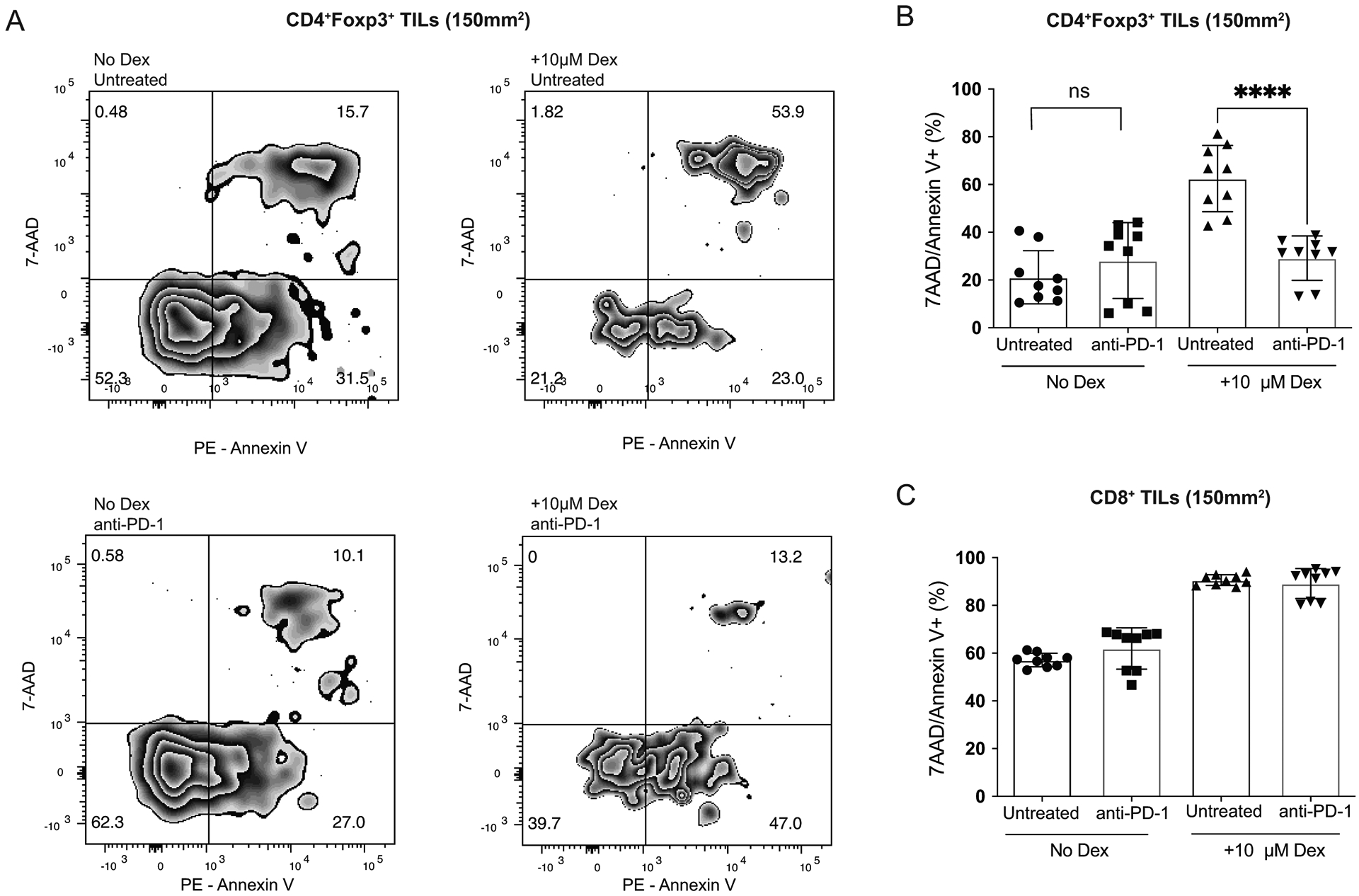
BALB/c Foxp3-GFP mice were injected with 1 × 10⁴ T11 (claudin-low) tumor cells. Mice were untreated or treated with 200 μg α-PD-1 antibody (J43) injected IP twice a week for the duration of the experiment. Tumors were harvested at 150mm2, digested, enriched for lymphocytes, and total T cells were isolated using cell isolation column (n=9). Isolated total T cells were cultured in 96 well plate in complete media or complete media + 10μM Dexamethasone. Apoptosis was measured using Annexin V and 7-AAD staining. (A) Representative flow plots gated on GFP+ Tregs isolated from the tumor of mice either untreated or treated with α-PD-1 cultured with or without Dex. (B) Percent CD4+Foxp3+7-AAD/Annexin V+ Tregs from CD45+ parent population. (C) Percent CD8+/7-AAD/Annexin V+ T cells from CD45+ parent population. Statistical significance determined by Mann-Whitney test. **** denotes p < 0.0001.